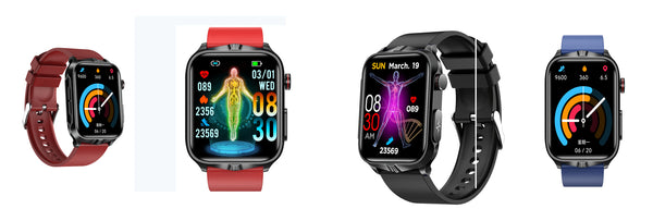
Non-invasive uric acid detection technology
- Near-infrared non-invasive uric acid detection technology
Gout is a common disease caused by abnormal levels of uric acid in the human body producing microcrystals, which can cause damage to human joints.
And microcrystals are not easily dissolved in human blood. In addition to slowing down body movements, they may also cause severe pain in human joints.
Pain and swelling. Based on its molecular structure and special chemical properties of infrared radiation, uric acid compounds can be detected in the infrared band.
things. Therefore, this article uses an infrared-visible spectrometer (UV-Vis spectrometer) to measure the concentration of artificial uric acid containing microcrystals.
concentration and absorption cross-section, and compared with uric acid extracted from human serum. Dissolve the uric acid sample in powder form with distilled water
The solution is converted into liquid form, starting with the uric acid raw material and serially diluting it to the other three concentrations based on molar concentration formula calculations.
The sample still contains coarse particles or suspended particles of uric acid that are not completely dissolved in the distilled water, which can be assumed to be artificial microcrystals to mimic
Actual microcrystals formed by gout.
The spectrometer (UV-VIS Maya 2000 Pro, Ocean Optics) was set to the UV wavelength region using a deuterium light source.
Prepare several plastic cuvettes for placing uric acid samples. Prepare by dissolving 100mg of uric acid powder in 15mL of distilled water
Uric acid stock solution (1.0mg/ml). Heat the mixture to 60°C on a hot plate and stir with a magnetic stirrer on the hot plate.
Then cool to 20-26˚ C. Prepare a uric acid stock solution by adding 85 mL of distilled water to the mixture to bring it to 100 mL. Dilute the original solution
into three different concentrations: 0.8mg/ml, 0.6mg/ml and 0.4mg/mL, placed in cuvettes respectively.
Figure 1 shows the complete experimental setup for the detection of uric acid, with the spectrometer connected to the computer via USB. Fiber optic from deuterium light source
Connect to the internal port of the cuvette holder and from the external port of the cuvette holder to the spectrometer. Before starting the experiment,
Heat the deuterium light source for about 20 seconds. Adjust the spectrometer integration time.
Calculation of uric acid absorption and concentration is based on Beer's Lambert Law formula, as shown in Equation 1.0. will transmit and
Substituting the incident light intensity into this equation, the value of the absorption cross-section can be calculated using the standard equation and deriving Equation 1.1.


Among them, A is the absorbance. ɛ is the molar absorption coefficient, a constant related to the chemical properties of the experimental sample. C is uric acid sample
product concentration. ɭ is the path length. Io is the incident light intensity. It is the transmitted light intensity. N is the molecular concentration (cm
3)
Uric acid detection is performed by analyzing the infrared light absorbance of uric acid compounds. Different concentrations of uric acid at the same wavelength have
There are different absorbance values. According to existing research results, the maximum absorption is obtained at 1000nm.
Figure 2 shows the same wave

Graph the initial results for four concentrations of uric acid.
Figure 2 Absorbance curves of uric acid at different concentrations
It can be seen from this absorbance curve that the uric acid stock solution with a concentration of 1 mg/mL has the maximum absorbance, and is then divided into
There are three other concentrations: 0.8mg/ml, 0.6mg/mL and 0.4mg/mL. The experiment successfully demonstrated that the spectrometer can perform ultraviolet
Different concentrations of uric acid are detected in the external band
